Using data science to achieve ultra-low dose CT image reconstruction
Ultra-low dose CT scans that provide superior image quality could not only benefit patients, but they could open up entirely new clinical applications.

 Enlarge
Prof. Jeff Fessler and Prof. Yong Long (PhD EE:Systems ’11) of the University of Michigan-Shanghai Jiao Tong University (UM-SJTU) Joint Institute are collaborating on a project to develop a dramatically improved approach to low-dose X-ray Computed Tomography (CT) image formation by extracting and using information from a big-data corpus of regular dose X-Ray CT images. The research is funded through the U-M/SJTU Collaborative Research Programs for Energy and Biomedical Technology, which funds projects that have future commercial potential.
CT provides high-resolution images of anatomical structures for diagnosis and management of human diseases and disorders. It has already had a tremendous impact on cancer. For example, CT data are used routinely to help detect abnormal growths, diagnose tumors, plan treatment for radiation oncology, guide biopsy procedures, assess of tumor responses to treatment and monitor for recurrence. Because of their value in patient care, there has been a dramatic increase in the number of CT scans ordered over the past several decades. In fact, CT scans are now are responsible for nearly 50% of the radiation exposure from medical imaging in the U.S. Unfortunately, the ionizing radiation in the form of X-rays used in CT scans is energetic enough to directly or indirectly damage DNA, putting patients at a higher risk for radiation-induced cancer. Studies have suggested that current CT usage may be responsible for 1.5%-2% of all cancers in the U.S.
Ultra-low dose CT (ULDCT) scans that provide superior image quality could not only benefit patients who currently rely on the technology, but they could open up entirely new clinical applications. For example, the U.S. Preventive Services Task Force recommends annual screening for lung cancer with LDCT in high-risk adults. The same screening could be available to a wider population of adults if ultra-LDCT scans were available. This would help with detection and treatment of early-stage lung cancer, and prevent a substantial number of lung cancer-related deaths, especially in China where lung cancer is the leading cause of cancer-related death. The death rate of lung cancer patients in China has increased by 465% over the past thirty years, and the number of individuals suffering from lung cancer in China could top one million by 2025.
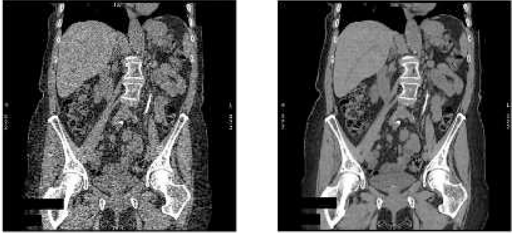
Currently most commercial CT scanners use a technique called filter-back projection (FBP) for image reconstruction. However, FBP requires higher than desired doses of radiation to produce high-quality diagnostic images. Model-based image reconstruction (MBIR) methods, also known as statistical image reconstruction methods, improve the ability to produce high-quality and accurate images, while greatly reducing patient exposure to potentially harmful levels of radiation. (see Fig. 1.). General Electrical introduced the world’s first commercial MBIR product, Veo, which allows CT scans to be performed using a radiation dose that is up to 75% lower than conventional image construction technique. It is currently in use at U-M Hospital.
To achieve an even greater reduction in the radiation to achieve ULDCT scans, Prof. Fessler and Prof. Long plan to take an entirely new approach, one that makes use of the vast archive of existing CT images of patients with similar ailments.

 Enlarge
Prior information extracted from big data collections of CT images should allow dramatic improvements in reconstructing high-quality images from ULDCT measurements. Motivated by the success of MBIR in lowering CT dose and the promising impact of big data on improving image quality of ULDCT imaging, this project aims to incorporate prior knowledge extracted from big data collections of clinical CT images into the design of the MBIR methods for X-ray CT. The proposed improved MBIR methods are expected to provide high image quality while further reducing the X-ray radiation dose to an ultra-low level.
Posted: August, 2015
 MENU
MENU 
